小而美-2019年尼康微观世界显微摄影大赛获奖作品
As scientists scour the natural world in search of truth, they often capture its beauty.
Nikon's Small World is regarded as the leading forum for showcasing the beauty and complexity of life as seen through the light microscope. ... The video competition, entitled Small World In Motion encompasses any movie or digital time-lapse photography taken through the microscope.
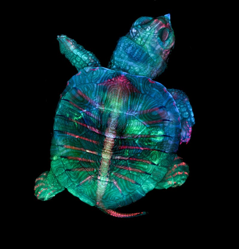
1st:
Fluorescent turtle embryo
location:Campbell Hall, New York, USA
Technique: Stereomicroscopy, Fluorescence
Magnification: 5x (Objective Lens Magnification)
More about this entry:
For microscopy technician Teresa Zgoda and recent university graduate Teresa Kugler, microscopy is a discipline that allows them to blend their dual passions of art and science. The 2019 winning image is a spectacular example of that, featuring a colorful turtle embryo captured using a combination of fluorescence and stereomicroscopy.
The pair captured this image while assisting in the Marine Biological Laboratory’s embryology course. It was here they learned the precise technique required to properly prepare various types of embryos to be observed and photographed. Creating this image was a unique challenge, largely due to the size of the sample. Over an inch long, and thick, it took time and precision to ensure the entire subject was photographed completely. What’s more, the magnification used meant only a small part of the turtle could be imaged on the focal plane. The final image is a compilation of hundreds of images that had to be stacked and stitched together.
When they are not taking photos through the microscope, both women enjoy being creative (for Kugler, that means cosplay, and for Zgoda it means photographing the landscapes, plants, and animals she sees on her hikes). Zgoda has recently started a job in a Boston hospital in a lab focused on neurology, while Kugler is excited to see what the world of science has in store for new Rochester Institute of Technology graduate.
“Microscopy lets us get a better look at the small things in life,” said Kugler, “It allows me to do science with a purpose.”
“We are inspired by the beautiful images we see through the microscope,” added Zgoda, “It’s amazing to be able to share that science with other people.”
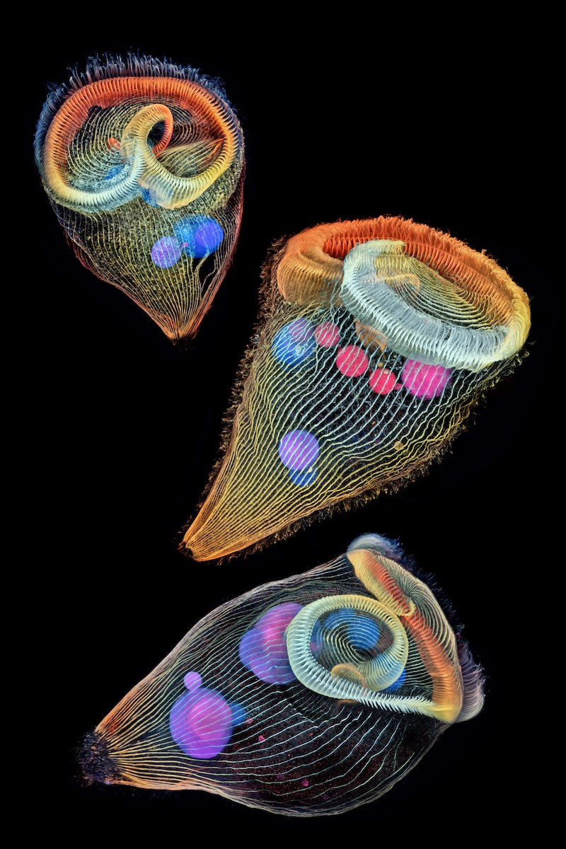
2nd:
Depth-color coded projections of three stentors (single-cell freshwater protozoans)
location:Janelia Research Campus Ashburn, Virginia, USA
Technique: unkown
Magnification: 40x (Objective Lens Magnification)
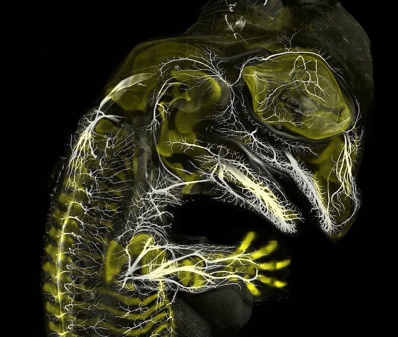
3rd:
Alligator embryo developing nerves and skeleton
location:Yale University
Technique: Immunofluorescence
Magnification: 10x (Objective Lens Magnification)
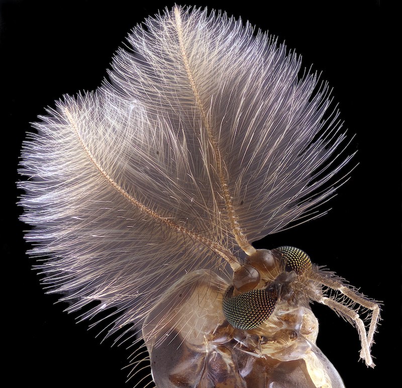
4th:
Male mosquito
location:University of Rostock Rostock, Mecklenburg Vorpommern, Germany
Technique: Focus Stacking
Magnification: 6.3x (Objective Lens Magnification)
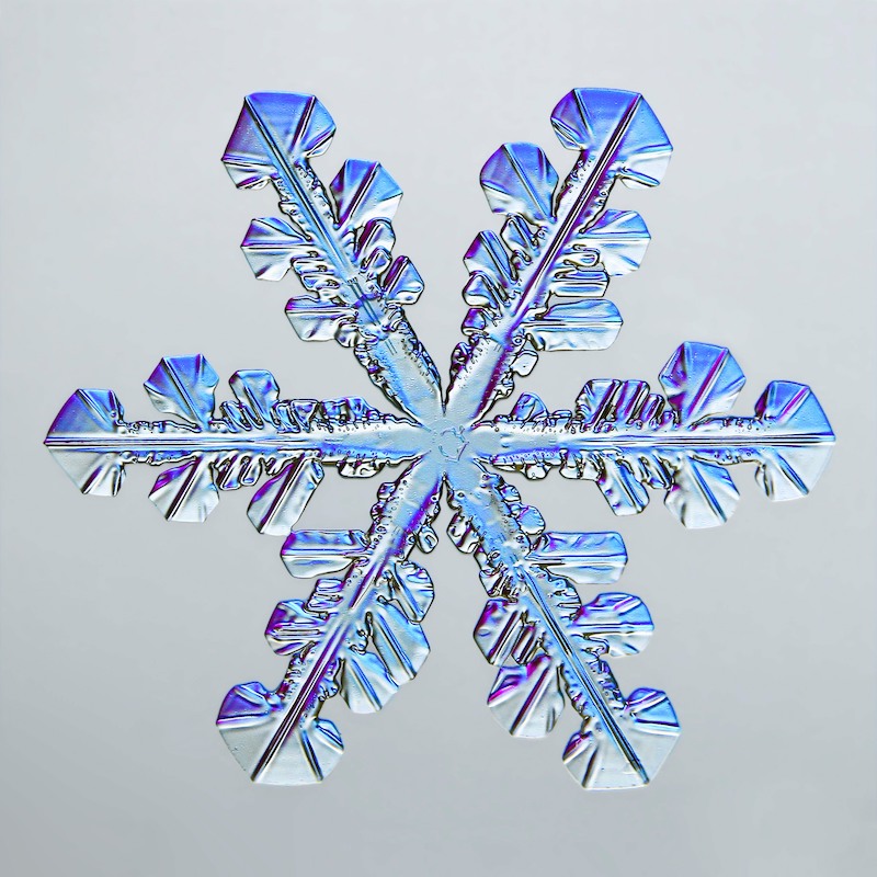
5th:
Snowflake
location:Caleb Foster Photography Jericho, Vermont, USA
Technique: Transmitted Light
Magnification: 4x (Objective Lens Magnification)
Visit more pictures click link: Nikon's small world