关于呼吸系统和我们如何进行呼吸
Respiratory System and How We Breathe
By Regina Bailey
 Credit: LEONELLO CALVETTI/Getty Images
The respiratory system is composed of a group of muscles, blood vessels, and organs that enable us to breathe. The primary function of this system is to provide body tissues and cells with life-giving oxygen while expelling carbon dioxide. These gases are transported via the blood to sites of gas exchange (lungs and cells) by the circulatory system. In addition to breathing, the respiratory system also assists in vocalization and the sense of smell.
Respiratory System Structures
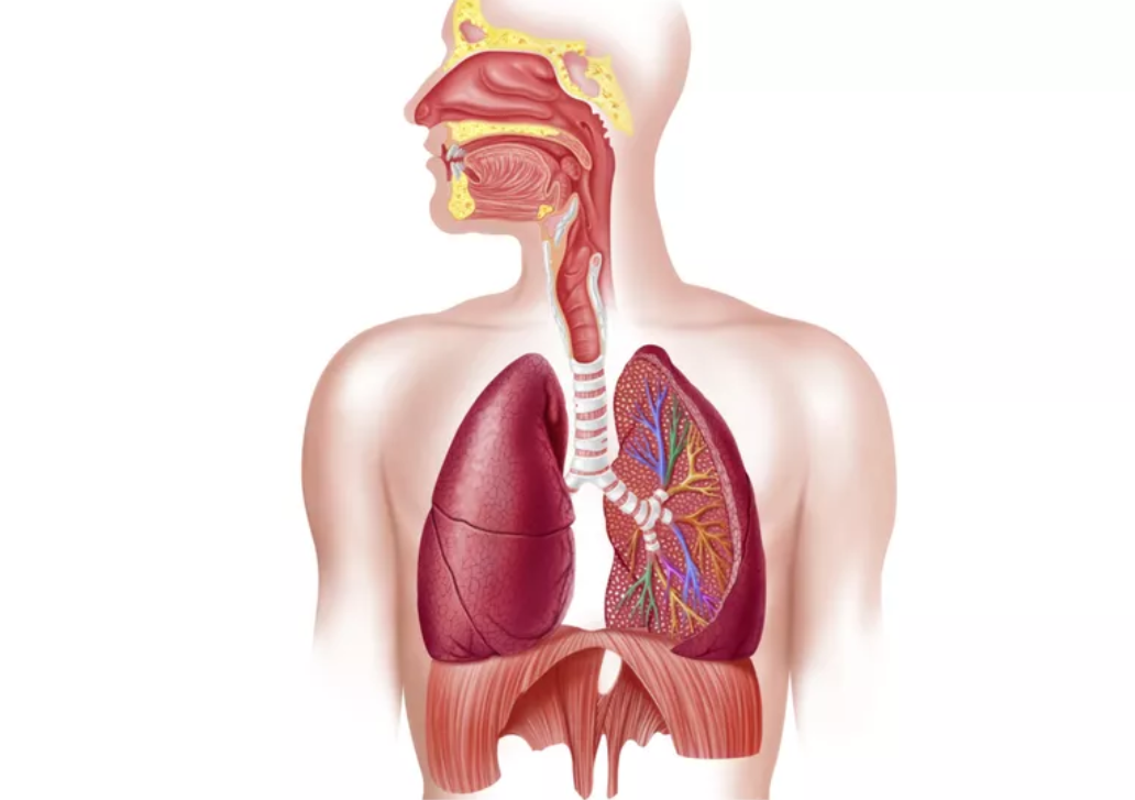
Respiratory system structures help to bring air from the environment into the body and expel gaseous waste from the body. These structures are typically grouped into three main categories: air passages, pulmonary vessels, and respiratory muscles.
Air Passages
- Nose and Mouth: openings that allow outside air to flow into the lungs.
- Pharynx (throat): directs air from the nose and mouth to the larynx.
- Larynx (voice box): directs air to the windpipe and contains vocal cords for vocalization.
- Trachea (windpipe): splits into left and right bronchial tubes that direct air to the left and right lungs.
Pulmonary Vessels
- Lungs: paired organs in the chest cavity that enable gas exchange between the blood and the air. The lungs are divided into five lobes.
- Bronchial tubes: tubes within the lungs that direct air into bronchioles and lets air out of the lungs.
- Bronchioles: smaller bronchial tubes within the lungs that direct air to small air sacs known as alveoli.
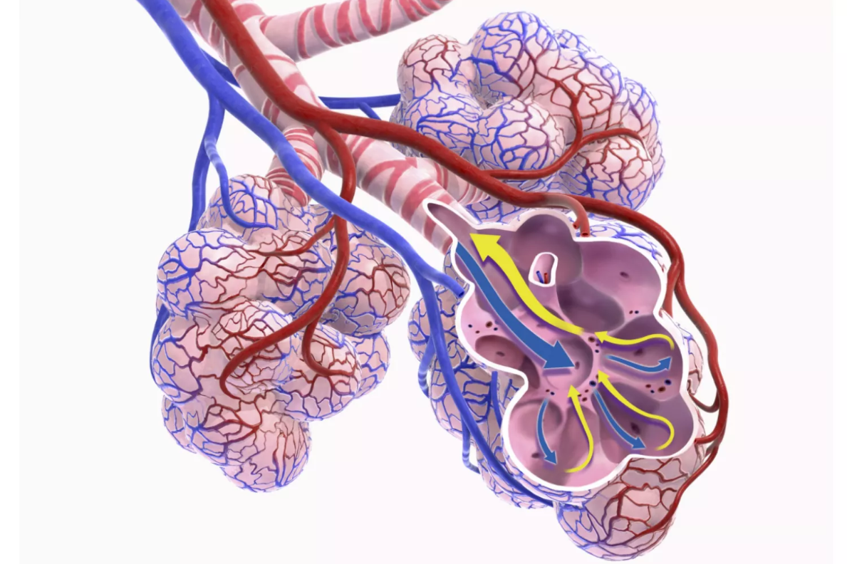
- Alveoli: bronchiole terminal sacs that are surrounded by capillaries and are the respiratory surfaces of the lungs.
- Pulmonary arteries: blood vessels that transport oxygen-depleted blood from the heart to the lungs.
- Pulmonary veins: blood vessels that transport oxygen-rich blood from the lungs back to the heart.
Respiratory Muscles
- Diaphragm: muscular partition that separates the chest cavity from the abdominal cavity. It contracts and relaxes to enable breathing.
- Intercostal muscles: several groups of muscles located between the ribs that help to expand and shrink the chest cavity to aid in breathing.
- Abdominal muscles: aid in faster exhalation of air.
How We Breathe
 Dorling Kindersley/Getty Images
Breathing is a complex physiological process that is performed by respiratory system structures. There are a number of facets involved in breathing. Air must be able to flow into and out of the lungs. Gases must be able to be exchanged between the air and blood, as well as between the blood and body cells. All of these factors must be under strict control and the respiratory system must able to respond to changing demands when necessary.
Inhalation and Exhalation
Air is brought into the lungs by actions of respiratory muscles. The diaphragm is shaped like a dome and is at its maximum height when it is relaxed. This shape reduces the volume in the chest cavity. As the diaphragm contracts, the diaphragm moves downward and the intercostal muscles move outward. These actions increase volume in the chest cavity and lower air pressure within the lungs. The lower air pressure in the lungs causes air to be drawn into the lungs through the nasal passages until the pressure differences equalize. When the diaphragm relaxes again, space within the chest cavity decreases and the air is forced out of the lungs.
Gas Exchange
Air is brought into the lungs from the external environment contains oxygen needed for body tissues. This air fills tiny air sacs in the lungs called alveoli. Pulmonary arteries transport oxygen-depleted blood containing carbon dioxide to the lungs. These arteries form smaller blood vessels called arterioles which send blood to capillaries surrounding millions of lung alveoli. Lung alveoli are coated with a moist film that dissolves air. Oxygen levels within the alveoli sacs is at a higher concentration than oxygen levels in the capillaries surrounding the alveoli. As a result, oxygen diffuses across the thin endothelium of the alveoli sacs into the blood within the surrounding capillaries. At the same time, carbon dioxide diffuses from the blood into the alveoli sacs and is exhaled through air passages. The oxygen-rich blood is then transported to the heart where it is pumped out to the rest of the body.
A similar exchange of gases takes place at body tissues and cells. Oxygen used by cells and tissues must be replaced. Gaseous waste products of cellular respiration such as carbon dioxide must be removed. This is accomplished through cardiovascular circulation. Carbon dioxide diffuses from cells into blood and is transported to the heart by veins. Oxygen in arterial blood diffuses from the blood into cells.
Respiratory System Control
The process of breathing is under the direction of the peripheral nervous system (PNS). The autonomic system of the PNS controls involuntary processes such as breathing. The medulla oblongata of the brain regulates breathing. Neurons in the medulla send signals to the diaphragm and the intercostal muscles to regulate the contractions which initiate the breathing process. The respiratory centers in the medulla control breathing rate and can speed up or slow down the process when needed. Sensors in the lungs, brain, blood vessels and muscles monitor changes in gas concentrations and alert respiratory centers of these changes. Sensors in air passages detect the presence of irritants such as smoke, pollen, or water. These sensors send nerve signals to respiratory centers to induce coughing or sneezing to expel the irritants. Breathing can also be influenced voluntarily by the cerebral cortex. This is what allows you to voluntarily speed up your breathing rate or hold your breath. These actions, however, can be overridden by the autonomic nervous system.
Respiratory Infection
 BSIP/UIG/Getty Images
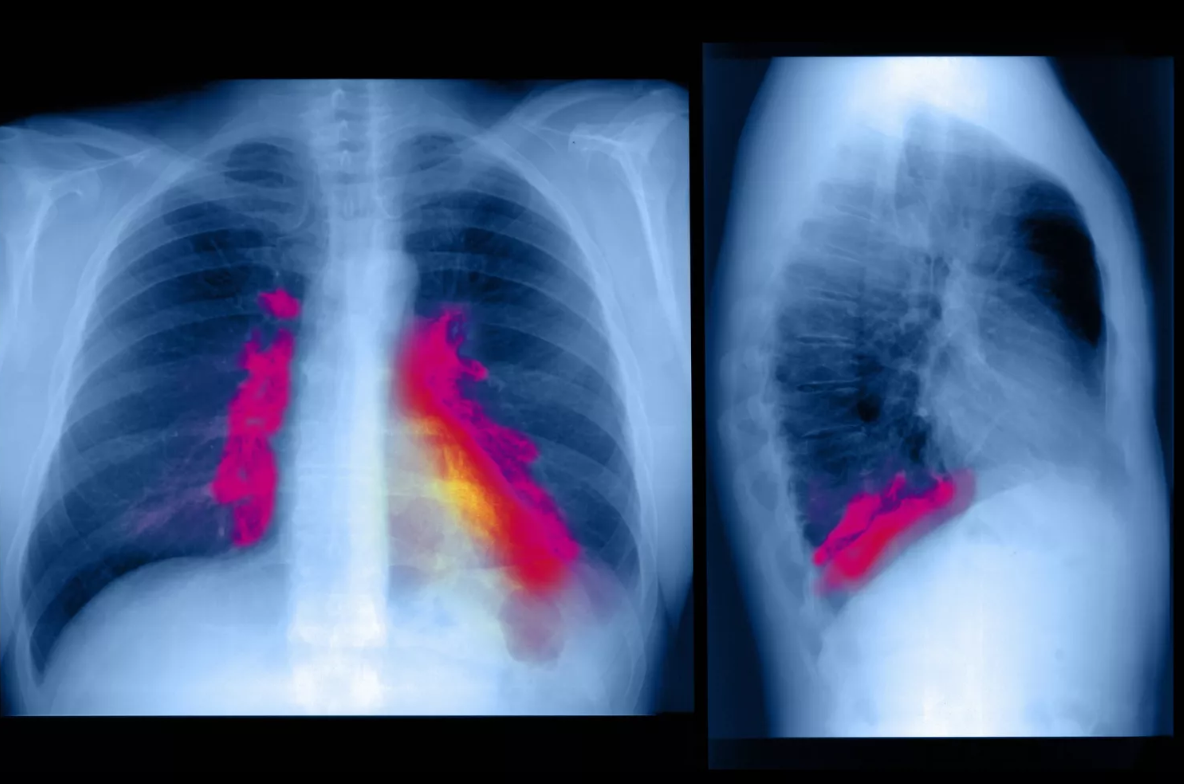
Respiratory system infections are common as respiratory structures are exposed to the external environment. Respiratory structures sometimes come in contact with infectious agents like bacteria and viruses. These germs infect respiratory tissue causing inflammation and can impact the upper respiratory tract as well as the lower respiratory tract.
The common cold is the most notable type of upper respiratory tract infection. Other types of upper respiratory tract infections include sinusitis (inflammation of the sinuses), tonsillitis (inflammation of the tonsils), epiglottitis (inflammation of the epiglottis that covers the trachea), laryngitis (inflammation of the larynx) and influenza.
Lower respiratory tract infections are often far more dangerous than upper respiratory tract infections. Lower respiratory tract structures include the trachea, bronchial tubes, and lungs. Bronchitis (inflammation of the bronchial tubes), pneumonia (inflammation of the lung alveoli), tuberculosis, and influenza are types of lower respiratory tract infections.
Key Takeaways
The respiratory system enables organisms to breathe. Its components are a group of muscles, blood vessels, and organs. Its primary function is to provide oxygen while expelling carbon dioxide.
Structures of the respiratory system can be grouped into three main categories: air passages, pulmonary vessels, and respiratory muscles.
Examples of respiratory structures include the nose, mouth, lungs, and diaphragm.
In the breathing process, air flows into and out of the lungs. Gases are exchanged between the air and blood. Gases are also exchanged between the blood and body cells.
All facets of breathing are under strict control as the respiratory system must be able to adapt to changing needs.
Respiratory system infections can be common since its component structures are exposed to the environment. Bacteria and viruses can infect the respiratory system and cause disease.